Home » Without Label » Blood Vessels Labeled / lesson 36: vessels and nerves of the abdomen & pelvis at ... - Around the t4 vertebra the aortic arch transitions into the thoracic aorta.
Blood Vessels Labeled / lesson 36: vessels and nerves of the abdomen & pelvis at ... - Around the t4 vertebra the aortic arch transitions into the thoracic aorta.
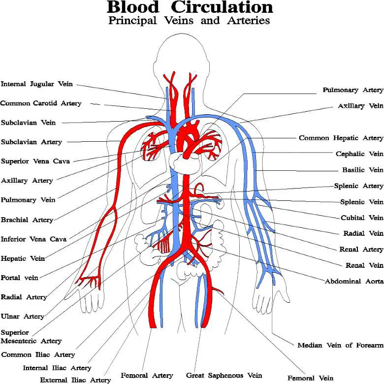
Blood Vessels Labeled / lesson 36: vessels and nerves of the abdomen & pelvis at ... - Around the t4 vertebra the aortic arch transitions into the thoracic aorta.. External veins and arteries of the heart ec by mrsdohm 64,633 plays 8p image quiz. Eventually, the smallest arteries, vessels called arterioles, further branch into tiny capillaries, where nutrients and wastes are exchanged, and then combine with other vessels that exit capillaries to form venules, small blood vessels that carry blood to a vein, a larger blood vessel that returns blood to the heart. Dr calum worsley and assoc prof craig hacking et al. The major arteries in the body. Around the t4 vertebra the aortic arch transitions into the thoracic aorta.
Blood vessels are the specially designed tubes that carry blood throughout the body. Blood vessel labeling 7p image quiz. Internal jugular vein • this is the larger of two vessels that drain blood from the head and neck into the subclavian. May 31, 2021 reading time: Dimitrios mytilinaios md, phd last reviewed:
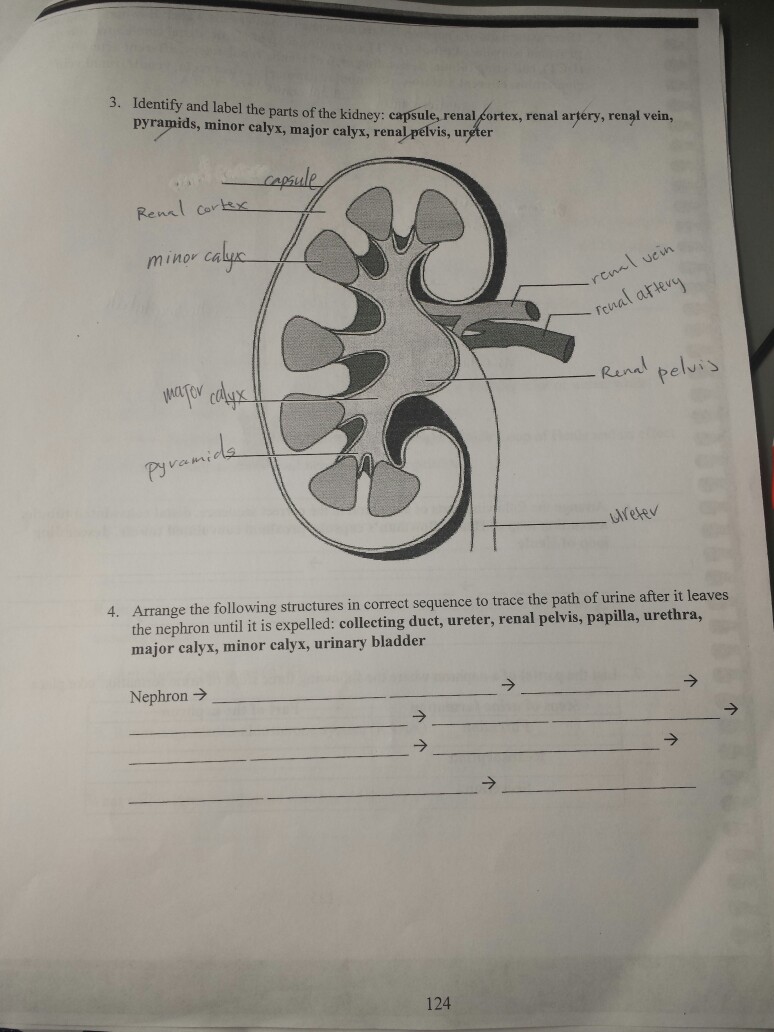
34 Label The Blood Vessels And Parts Of The Nephron ... from d2vlcm61l7u1fs.cloudfront.net This video series covers the blood vessels for anatomy and physiology ii students. The common cartoid artery extends from the brachiocephalic artery. The aorta has three crucial segments. Blood vessel labeling 7p image quiz. Use key choices to identify the blood vessel tunic described. The deep brachial artery of the arm. The adventitia or outer layer which provides structural support and shape to the vessel Vessels transport nutrients to organs/tissues and to transport wastes away from organs/tissues in the blood.
Very small branches that collect the blood from the various organs and parts are called venules, and they unite to form veins, which return the blood to the heart.
Three blood vessels branch from the aortic arch: Blood vessels 11p image quiz. Tunica intima, tunica media, tunica externa tunica media, tunica intima, tunica externa tunica externa, tunica intima, tunica media The function and structure of each segment of the peripheral vascular system vary depending on the organ it supplies. Around the t4 vertebra the aortic arch transitions into the thoracic aorta. Blood cells by descartes 48,654 plays 9p image quiz. Bulky middle tunic contains smooth muscle and elastin 3. Blood vessels anatomy blood vessels are responsible for the transportation of blood, made up arteries and veins, they creates pathways for the oxygenated blood to travel to their destination and pathways for the used deoxygenated blood to travel back to the heart or lungs.capillaries are designed to permit the transfer of gasses within the blood, such as the delivery of oxygen and the return. The word vascular, meaning relating to the blood vessels, is derived from the latin vas, meaning vessel. Lorenzo crumbie mbbs, bsc • reviewer: Blood vessel labeling 7p image quiz. The inferior vena cava is labeled in the figure below. The brachiocephalic artery, the left subclavian artery, and the left common carotid artery.
The ulnar artery of the forearm. It extends on each side of the neck and divides at the level of the larynx into two branches: Blood vessel labeling 15p image quiz. Describe the development of blood vessels and fetal circulation; The main blood supply of the eye arises from the ophthalmic artery, which gives off orbital and optical group branches.
Blood vessels diagram from healthiack.com •formed where capillaries unite • extremely porous 1) venules: The deep brachial artery of the arm. The tunica externa, the tunica media, and the tunica intima. The left ventricle of the heart pumps oxygenated blood into the aorta. Veins (in blue) are the blood vessels that return blood to the heart. Deep veins, located in the center of the leg near the leg bones, are enclosed by muscle. Like arteries, veins form a complex, branching system of larger and smaller vessels. Place in order from superficial to deep the three tunics of a typical blood vessel.
Aside from capillaries, blood vessels are all made of three layers:
Veins (in blue) are the blood vessels that return blood to the heart. The cephalic artery of the arm. The tunica externa, the tunica media, and the tunica intima. Anatomy of blood vessels review sheet 32 261 microscopic structure of the blood vessels 1. The vessels that carry blood away from the heart are called arteries, and their very small branches are arterioles. Bonner walks through the dissection of a cat's blood vessels. The deep brachial artery of the arm. Blood vessels of the head and neck. 4 but is clearly visible entering the right atrium of the heart. Blood vessel labeling 9p image quiz. Blood vessels are the specially designed tubes that carry blood throughout the body. The word vascular, meaning relating to the blood vessels, is derived from the latin vas, meaning vessel. Blood vessel labeling 7p image quiz.
While most blood vessels are located deep from the surface and are not visible, the. Navigate to the cardiovascular system area in the following pal 3.0 module: It extends on each side of the neck and divides at the level of the larynx into two branches: Around the t4 vertebra the aortic arch transitions into the thoracic aorta. The main blood supply of the eye arises from the ophthalmic artery, which gives off orbital and optical group branches.
torso model anatomy labeled 434903 orig - Top Label Maker from labels-top.com Label the major blood vessels of the pulmonary and systemic circulations; Vessels transport nutrients to organs/tissues and to transport wastes away from organs/tissues in the blood. Blood supply of the head and neck alila medical images. Very small branches that collect the blood from the various organs and parts are called venules, and they unite to form veins, which return the blood to the heart. Blood vessels labeled head : This article lists a series of labeled imaging anatomy cases by system and modality. Structure & function of blood vessels. 15 minutes it would be impossible to get blood to the predestined locations without the vascular pathways.
Three blood vessels branch from the aortic arch:
Oxygenated blood flows away from the heart through arteries. The axillary artery of the arm. Identify and describe the hepatic portal system; Dr calum worsley and assoc prof craig hacking et al. Dimitrios mytilinaios md, phd last reviewed: Human cadaver, anatomical models, histology, cat, and fetal pig. The superior vena cava is not labeled in figure 7.4. Veins (in blue) are the blood vessels that return blood to the heart. The aorta has three crucial segments. The main blood supply of the eye arises from the ophthalmic artery, which gives off orbital and optical group branches. Blood vessel labeling 7p image quiz. •formed where capillaries unite • extremely porous 1) venules: This article covers the anatomy, function and clinical relevance of the vessels and.